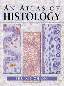
An Atlas of Histology
Shu-Xin Zhang
Mar 2013 · Springer Science & Business Media
5.0star
2 reviewsreport
Ebook
426
Pages
reportRatings and reviews aren’t verified Learn More
About this ebook
The beginning student of histology is frequently confronted by a paradox: diagrams in many books that illustrate human microanatomy in a simplified, cartoon-like manner are easy to understand, but are difficult to relate to actual tissue specimens or photographs. In turn, photographs often fail to show some important features of a given tissue, because no individual specimen can show all of the tissue's salient fea tures equally well. This atlas, filled with photo-realistic drawings, was prepared to help bridge the gap between the simplicity of diagrams and the more complex real ity of microstructure. All of the figures in this atlas were drawn from histological preparations used by students in my histology classes, at the level of light microscopy. Each drawing is not simply a depiction of an individual histological section, but is also a synthesis of the key structures and features seen in many preparations of similar tissues or organs. The illustrations are representative of the typical features of each tissue and organ. The atlas serves as a compendium of the basic morphological characteristics of human tissue which students should be able to recognize.
Ratings and reviews
5.0
2 reviews
Rate this ebook
Tell us what you think.
Reading information
Smartphones and tablets
Install the Google Play Books app for Android and iPad/iPhone. It syncs automatically with your account and allows you to read online or offline wherever you are.
Laptops and computers
You can listen to audiobooks purchased on Google Play using your computer's web browser.
eReaders and other devices
To read on e-ink devices like Kobo eReaders, you'll need to download a file and transfer it to your device. Follow the detailed Help Center instructions to transfer the files to supported eReaders.